Krystle C. Kalafut
Hepatic mTORC1 and nutrient signalling
Department of Molecular Metabolism, Harvard T.H. Chan School of Public Health · Department of Cell Biology, Harvard Medical School
Lab lineagetrained under Brendan D. Manning →
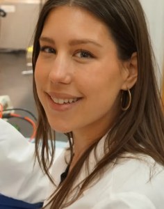 Portrait: Harvard T.H. Chan School of Public Health
Krystle C. Kalafut works in Brendan Manning's laboratory, jointly in the Department of Molecular Metabolism at the Harvard T.H. Chan School of Public Health and the Department of Cell Biology at Harvard Medical School.
She is first author on a 2026 Nature Communications study using liver-specific mouse genetic models to show that AKT-mediated TSC2 phosphorylation drives insulin's induction of hepatic mTORC1 but is dispensable for feeding-induced activation — which instead requires dietary protein — and that hepatic mTORC1 signalling is not elevated in diet-induced obesity despite impaired insulin and glucose homeostasis.
Milestones in the Atlas
A selection, not a full bibliography: only papers that meet the Atlas inclusion criteria.
| Year | Evidence | Study |
|---|---|---|
| 2026 | A | Dietary protein governs the role of insulin signaling in the postprandial regulation of hepatic mTORC1. KAL2026 First author showing hepatic mTORC1's postprandial activation depends on dietary protein, dissecting it from insulin/AKT/TSC2 signalling in liver-specific mouse models. |
| 2026 | A | Feeding-Induced Muscle mTORC1 Signaling Regulates Postprandial Protein Synthesis and Endurance but Not Muscle Size LAP2026 Using a mouse model expressing an AKT-nonphosphorylatable TSC2 mutant specifically in skeletal muscle, the Manning lab genetically separated feeding-induced from contraction-induced mTORC1 activation. AKT-mediated TSC2 phosphorylation is required for feeding (but not contraction) to activate muscle mTORC1 and drive postprandial protein synthesis — yet mice lacking this feeding-induced signal have normal muscle mass and myofiber size, and instead show improved maximal endurance capacity with a modest rise in mitochondrial content. The finding dissociates mTORC1's role in postprandial anabolism from its role in maintaining steady-state muscle mass, and is a directly relevant data point for whether the pattern/source of mTORC1 activation (feeding vs. mechanical) — not just its average level — shapes downstream outcomes. |
Co-authors in the Atlas
People with a profile here who share at least one study with Krystle C. Kalafut.
- Brendan D. Manning 2 shared studies Was among the groups that in 2002 identified TSC2 as a direct target of Akt, linking insulin and PI3K signalling to mTORC1, and later found TBC1D7 as the third subunit of the TSC complex. His lab has shown how mTORC1 turns growth signals into the synthesis of lipids and nucleotides
- Karen E. Inouye 2 shared studies Metabolic physiologist who runs the Hotamışlıgil lab’s physiology facility, which specialises in glucose clamps, and co-authored two 2026 mouse studies on how feeding controls mTORC1 in liver and muscle.
- Madi Y. Cissé 2 shared studies Research associate in the Manning lab and co-author on the two 2026 mouse studies showing which feeding signals switch on mTORC1 in liver and in skeletal muscle.
- Samuel C. Lapp 2 shared studies First author of the muscle study showing that feeding-induced mTORC1 activation through AKT–TSC2 drives postprandial protein synthesis but is not needed to build or keep muscle mass.
- Yann Cormerais 2 shared studies Research scientist in the Manning lab who works on how PI3K–AKT–TSC2 signalling controls mTORC1 in living tissues, using mouse models in which TSC2 cannot be phosphorylated by AKT.
- Gökhan S. Hotamışlıgil Leads a Harvard laboratory on metabolic disease and was elected to the US National Academy of Sciences in 2026 for achievements in metabolic research
Every researcher with a profile →
Thank you. Your answer was counted anonymously.
Thank you. That helps us know which pages have been read by someone who knows the work.
Thank you. We will check it and correct the page.