James H. Hurley
Structural biologist who uses cryo-EM to show how protein machines assembled on membranes control autophagy and nutrient signalling. His lab solved structures of the Rag–Ragulator complex and of the whole mTORC1–TFEB–Rag–Ragulator assembly on the lysosome
Professor of Molecular & Cell Biology and Kirsch Springer Chair in Biological Sciences, UC Berkeley (since 2013) · Faculty Scientist, Lawrence Berkeley National Laboratory (since 2014) · Chief, Section on Structural Biology & Cell Signaling, NIDDK/NIH (1998–2013) · Senior Investigator, NIDDK/NIH (1997–2013) · Tenure-track Investigator, NIH (1992–1997) · Postdoctoral Fellow, University of Oregon (1990–1992) · PhD in Biophysics, UCSF (1990) · BA in Physics, San Francisco State University (1984) · Member, US National Academy of Sciences (2020) · Hans Neurath Award, Protein Society (2014)
Hurley Lab, UC Berkeley (Berkeley, California, USA) ↗ Bluesky@jimhurley.bsky.social ↗ ORCID0000-0001-5054-5445 ↗
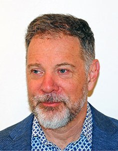 Portrait: UC Berkeley
mTORC1 only switches on when it is physically dragged to the surface of the lysosome, and the machine that does the dragging is a five-protein clamp called Ragulator together with the Rag GTPases. In the 2017 study, Hurley's laboratory solved the crystal structure of the whole human Ragulator at 1.4 angstrom resolution, which is sharp enough to see individual side chains. Lamtor1 turned out to wrap around the other four subunits like a belt, while the Lamtor2-Lamtor3 pair stacks on Lamtor4-Lamtor5 to build the platform the Rags sit on.
A crystal structure alone could not show where the Rags actually dock, so the team added two other methods. Hydrogen-deuterium exchange, which reports how exposed each stretch of protein is to water, mapped the Rag contact to the outer face of Lamtor2-Lamtor3 and to the floppy N-terminal tail of Lamtor1; electron microscopy then gave a lower-resolution picture of full-length RagA-RagC bound to Ragulator. Stitching these together produced a hybrid model in which the business ends of the Rags project away from the Ragulator core, held out like a handle for mTORC1 to grab.
Hurley spent two decades at the NIH before moving to Berkeley in 2013, and built his reputation on structures of the protein complexes that bend and cut membranes, especially the ESCRT machinery. His group now uses cryo-electron microscopy and cryo-electron tomography on the autophagy initiation machinery, on lysosome biology in cancer and Alzheimer's disease, and on how HIV hijacks membrane trafficking. The Ragulator work sits at the junction of those interests: it is a nutrient sensor, a membrane anchor and a drug-relevant target at the same time.
Milestones in the Atlas
| Year | Evidence | Study |
|---|---|---|
| 2017 | M | Hybrid Structure of the RagA/C-Ragulator mTORC1 Activation Complex SUX2017 Senior author (with Roberto Zoncu); his laboratory determined the 1.4 angstrom Ragulator crystal structure and combined it with hydrogen-deuterium exchange and EM into the hybrid RagA/C-Ragulator model. |
| 2023 | M | Structure of the lysosomal mTORC1-TFEB-Rag-Ragulator megacomplex CUI2023 First structural view of how mTORC1 actually reaches TFEB to phosphorylate it: two full Rag-Ragulator complexes present a single TFEB molecule to the mTOR active site, one in the normal Raptor-docking arrangement and a second, non-canonical one that grips TFEB's own first helix. Mutating that grip point drives TFEB straight into the nucleus (turning on autophagy/lysosome genes) without disturbing where mTORC1 itself sits. Explains, at the level of atoms, why TFEB phosphorylation needs the tumour suppressor FLCN and the GDP-loaded state of RagC in a way no other mTORC1 substrate does. Boundary: cryo-EM of a complex reconstituted from purified human proteins — a static structural snapshot, not a live cell or a real-time measurement. |
| 2025 | M | Structural basis for mTORC1 activation on the lysosomal membrane CUI2025 Resolves how mTORC1 flips from 'parked on the lysosome' to 'catalytically switched on'. Reconstituting the whole assembly on membranes from purified Rheb, Rag GTPases, Ragulator and mTORC1, cryo-EM shows the kinase reaches full activity only after two separate anchoring steps: Rag-Ragulator first pulls it to within about 100 angstroms of the membrane, then Rheb pulls it to within about 40 angstroms, and only when Raptor and mTOR itself also touch the membrane directly do the catalytic residues line up for full activity. Growth-factor input (via Rheb) and nutrient input (via the Rags) are shown converging on the same physical docking event rather than acting through separate switches. Boundary: reconstituted on synthetic membranes from purified components — a structural/biochemical model, not a measurement inside a living cell. |
Co-authors in the Atlas
People with a profile here who share at least one study with James H. Hurley.
- Andrea Ballabio 2 shared studies Discovered that the transcription factor TFEB is a master switch for building lysosomes and for autophagy. His lab was one of the groups that showed in 2012 that mTORC1 keeps TFEB out of the nucleus from the lysosome surface, and later (2020) that constitutive TFEB activation drives the kidney disease of Birt-Hogg-Dubé syndrome
Every researcher with a profile →
On the programme
Meetings in the Atlas calendar where James H. Hurley is listed among the speakers or organisers.
- past Keystone: Autophagy – Mechanisms to Therapy 2026 ↗ 2–5 Feb 2026 · Keystone, Colorado, USA · ★★